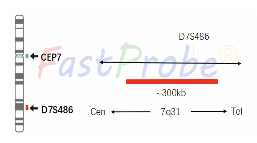
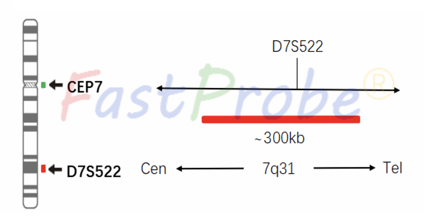
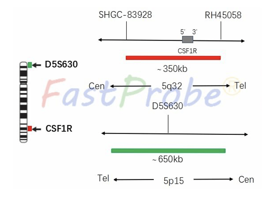
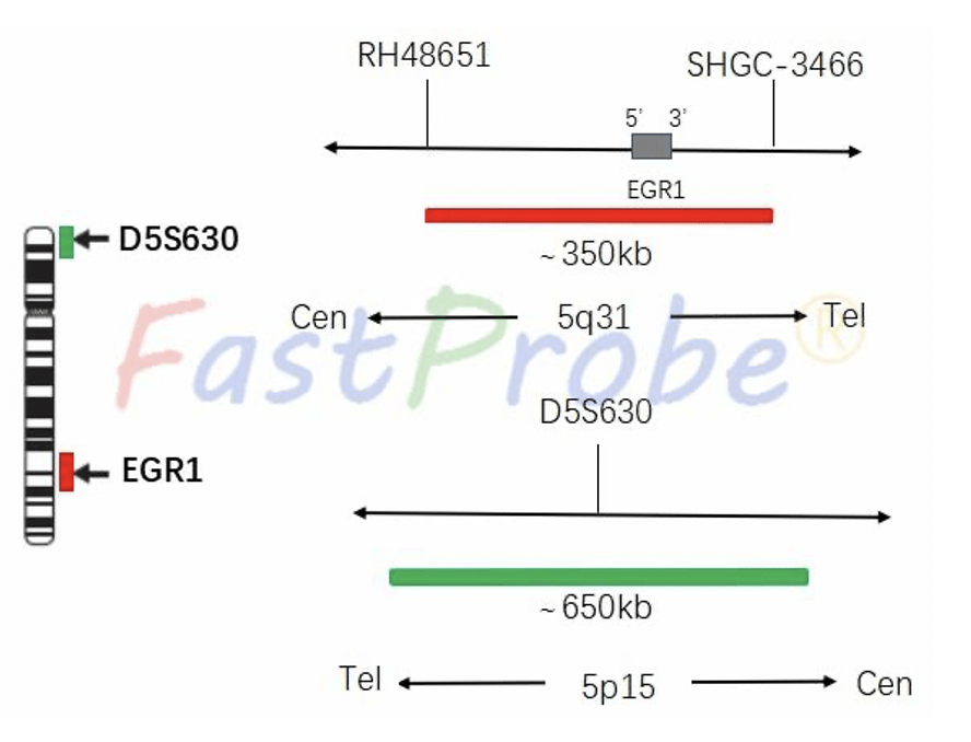
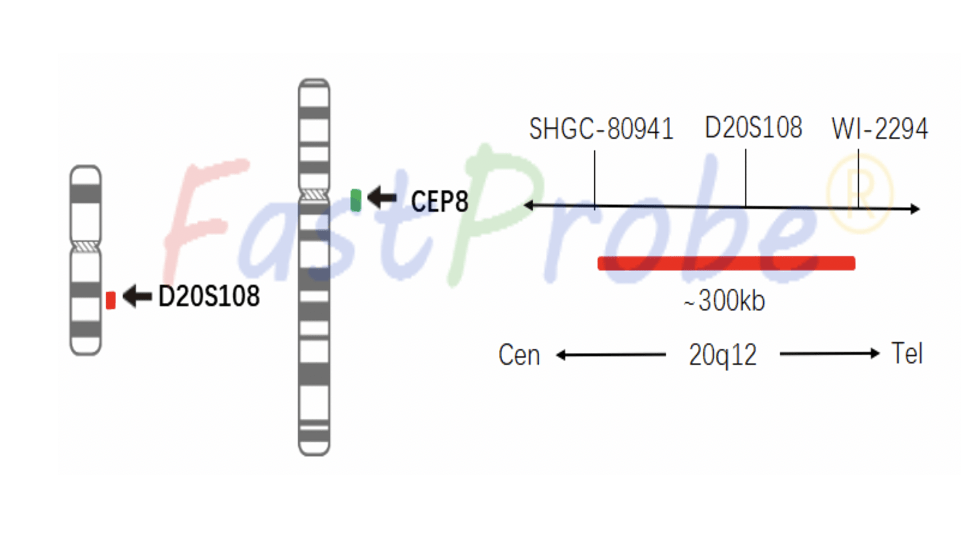
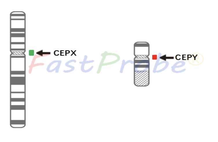
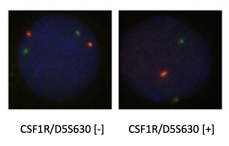
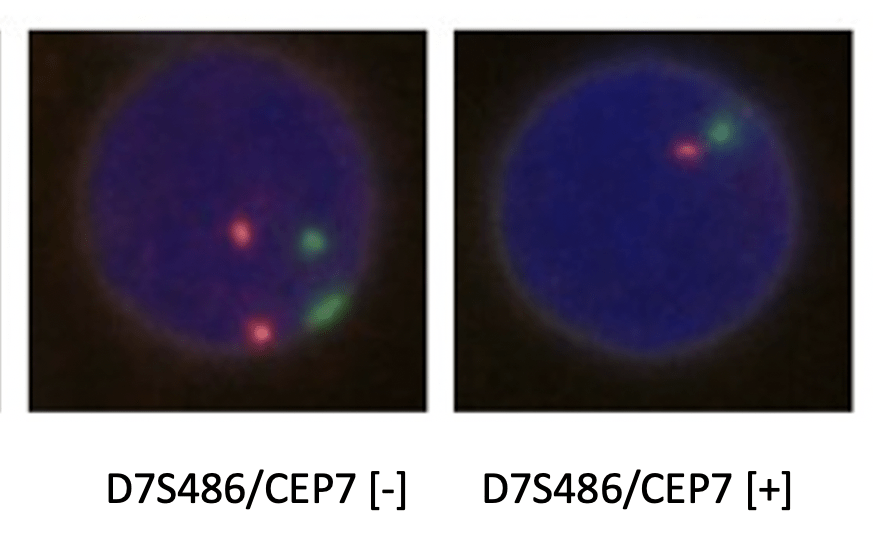
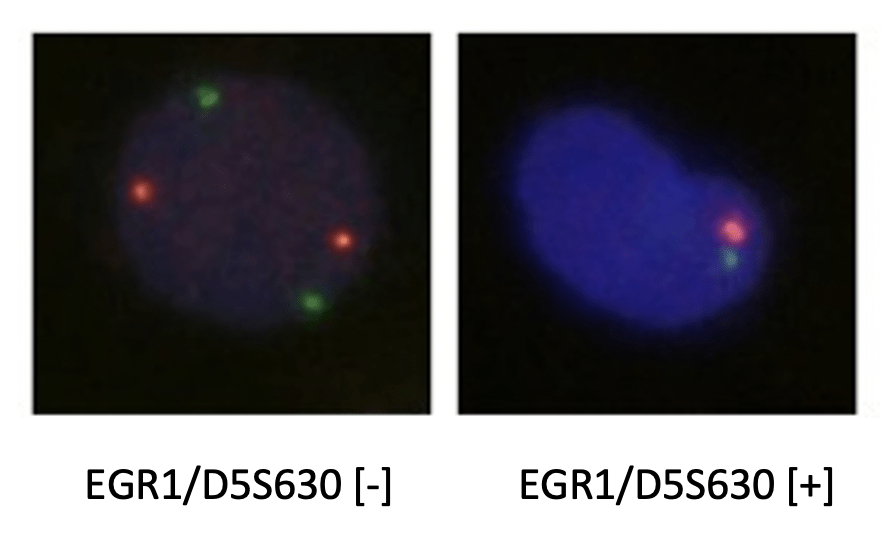
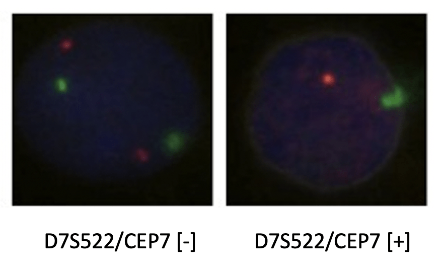
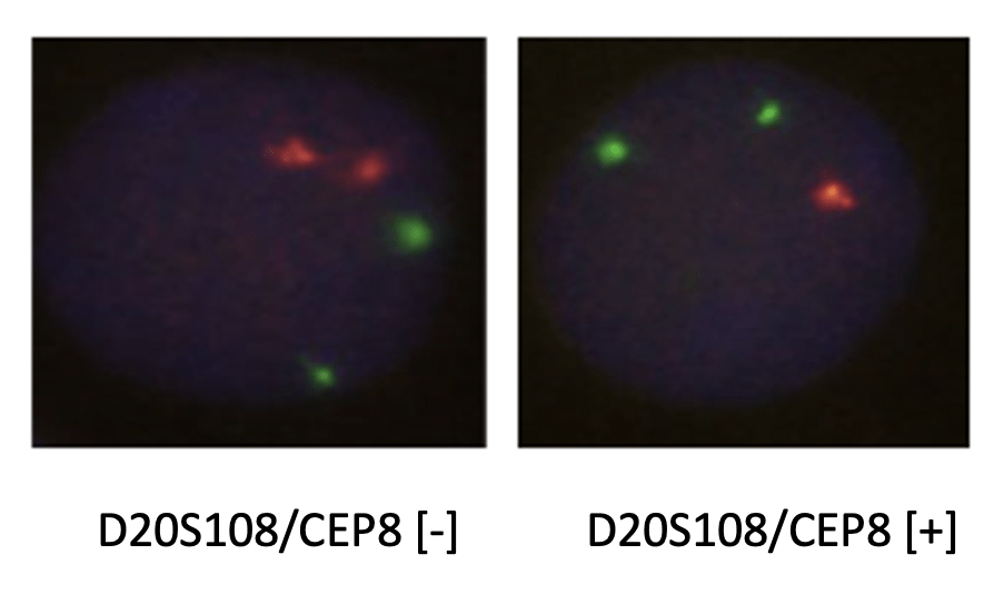
This kit uses an orange-red fluorescent dye to label CSF1R, EGR1, D7S486, D7S522, D20S108, CEPY probes, and a green fluorescent dye to label D5S630, CEP7, CEP8 and CEPX probes. The probes bind to the target detection site by in situ hybridization. Under normal conditions (no gene deletion and chromosome abnormality), two orange-red signals and two green signals are shown under a fluorescence microscope. When there is a gene deletion, there will be a lack of green or orange-red signal, and when there is a chromosomal polysomy, the centromere gene probe signal will increase. The detection of gene deletion and chromosome abnormality by FISH method is of great clinical significance for the diagnosis, treatment and prognosis of MDS.